

Vertebral Motion Analysis for Spinal Pain & Injury
Get Precise Answers With Advanced Real-Time Guided Spinal Motion Imaging.
Discover injuries that will never be picked up by a traditional X-ray or MRI. Schedule your consultation by clicking on one of the options below:
Don't Wait for Your Pain to Get Worse
Get back to doing the things you love—with answers and a clear path to recovery.
At HyperCharge Health, we give you answers when others can't.
-

Accident Injuries
We provide objective findings to get you the compensation you deserve.
-

Cervical Scan
We provide the only guided motion imaging in the state of Minnesota.
-
Lumbar Scan
We provide real-time spinal motion imaging that reveals hidden instabilities often missed by standard MRIs and X-rays.
The HyperCharge Health Difference: A Systems Approach to Nerve Health
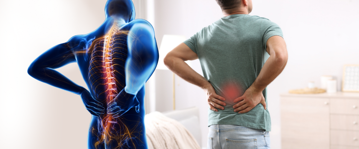
You've been living with persistent neck or back pain, perhaps after a car accident, a whiplash injury, or from years of degenerative changes. You've had the MRIs, the X-rays, and maybe even injections, yet you're told "everything looks normal" while your pain and instability persist. This experience is incredibly frustrating and leaves you without a clear path forward. The problem is that standard imaging takes a static snapshot, completely missing the injuries that only appear when you move. At HyperCharge Health in Edina, MN, we want you to know there is an answer. We use a revolutionary diagnostic tool called Vertebral Motion Analysis (VMA) to create a "motion picture" of your spine, revealing the hidden instabilities that are the true source of your pain and providing the objective data needed to finally heal.
Our Musculoskeletal Program and Institute for Regenerative Medicine is co-directed by our founder, a board-certified orthopedic spine surgeon, and Dr. Louis Saeger, a renowned triple board-certified pain management specialist. This dual-expert leadership is unmatched in Minnesota. Both physicians utilize VMA technology to solve complex cases of neck and back pain that have failed conventional diagnosis. The VMA scan is vital in their practices, providing the precise data needed to make critical treatment decisions—whether that means guiding one of Dr. Saeger's advanced regenerative procedures, determining the need for surgical intervention, or designing a non-invasive, integrative recovery plan. This allows us to target the root cause of instability with unparalleled precision.

Dr. Louis C. Saeger
You Deserve Better.
HyperCharge is Hope.

Why HyperCharge for VMA in Minnesota?
Dual-Expert Leadership: Your evaluation is overseen by both a board-certified orthopedic spine surgeon and a triple board-certified pain management specialist.
Radiation-Safe Dynamic Imaging: VMA uses advanced, low-dose fluoroscopy to safely visualize your spine in motion.
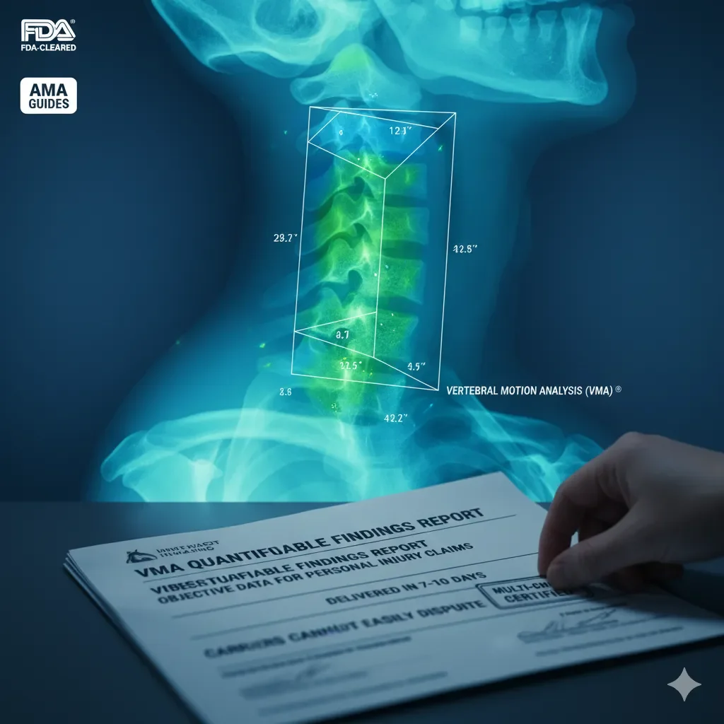
Objective Data for Treatment & Legal Cases: We provide court-admissible, quantitative proof of injury for motor vehicle and workers' comp claims.
Integrative Treatment Synergy: We are the only clinic in the Twin Cities that combines VMA diagnostics with a full suite of regenerative and biohacking therapies.
Specialized Imaging for Injury Diagnosis
We provide advanced scans for concussions, ligament injuries, and chronic spine conditions.
-
Cervical Scan
-
Lumbar Scan
Attorneys: Optimize Your PI Case Outcomes with the VMA®
-
The Problem
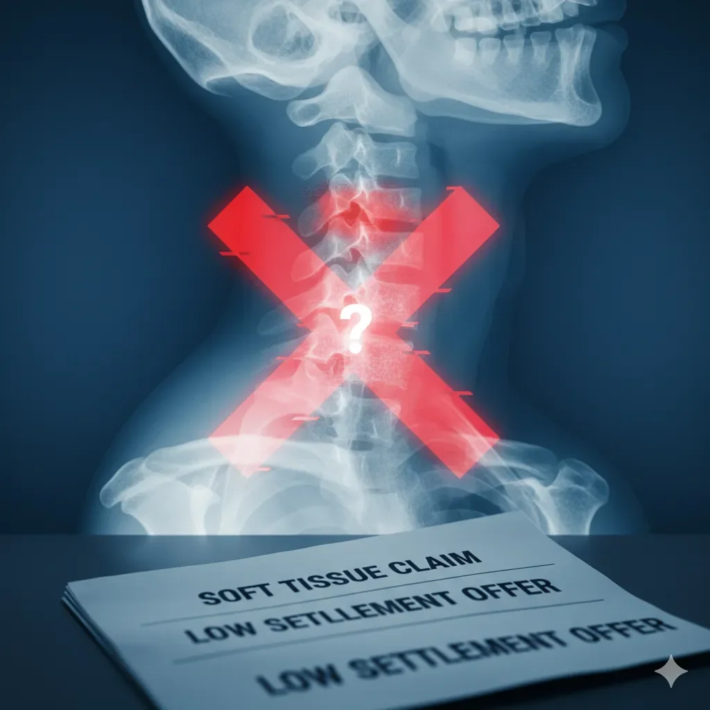
If you’ve handled personal injury cases, you already know the frustration: Carriers dismiss your claims as “soft tissue.” Standard imaging misses critical injuries. Settlement offers stay low, leaving your clients undercompensated.
-
The Solution
We provide Vertebral Motion Analysis (VMA) ®, an FDA-cleared and patented technology validated in the AMA Guides. VMA reveals spinal ligament injuries and other hidden conditions that X-rays and MRIs often miss.
The result? Quantifiable, objective findings that carriers cannot easily dispute. Reports are reviewed through a certified, multi-check process and typically delivered to providers within 7-10 business days.

Our Comprehensive VMA-Guided Toolkit
Explore how VMA works and how we integrate its findings into a powerful, multi-modal recovery plan.
-
Why Your MRI or X-Ray Missed the Problem
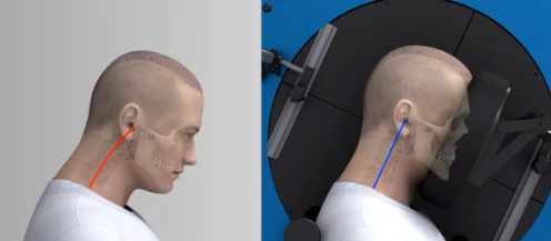
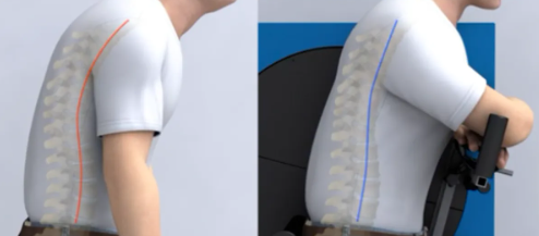
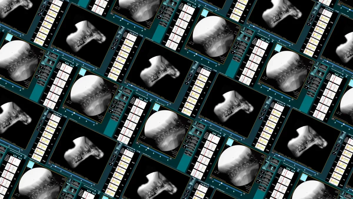
What It Is: Vertebral Motion Analysis (VMA) is a dynamic, video-based imaging tool that analyzes how your vertebrae move in relation to each other during controlled motion.
How It Helps: Standard MRIs, CT scans, and static X-rays are like a single photograph of your spine. They are excellent for showing structural problems like a herniated disc or a fracture, but they completely miss injuries to ligaments and other soft tissues that cause instability only when you move. VMA is like a motion picture, capturing these dynamic abnormalities in real-time.
How It Works (The Science): VMA uses AI-assisted, low-dose fluoroscopy to record your spine at 8 frames per second while a device gently guides you through flexion and extension. Computerized tracking then precisely measures the translation and rotation between each vertebra, identifying segmental instability with up to 50% less measurement variability than old flexion-extension X-rays.
Important Safety Information: VMA uses a low dose of radiation, approximately 25% less than a traditional flexion-extension X-ray series, and is considered very safe. It is contraindicated during pregnancy.
-
Objective, Court-Admissible Proof of Injury
What It Is: A specialized application of VMA to provide quantitative, objective evidence for personal injury and workers' compensation cases.
How It Helps: For patients injured in motor vehicle accidents or work-related incidents, VMA is indispensable. It provides objective, court-admissible data that can strengthen your case by proving the existence and severity of a ligamentous injury. This can elevate a diagnosis from a simple "strain/sprain" to a demonstrable, permanent impairment, which can significantly increase case value and ensure you receive appropriate compensation for your injury.
How It Works (The Science): By precisely quantifying abnormal vertebral translation, VMA provides measurable proof of ligamentous laxity or damage. Standard imaging often misses these injuries up to 80% of the time, but VMA can document them with up to 41% greater specificity than traditional methods, providing your legal team with the hard evidence they need.
-
How VMA Personalizes Your HyperCharge Protocol
What It Is: The process of using the specific findings from your VMA scan to design a targeted, multi-modal recovery plan.
How It Helps: VMA doesn't just diagnose the problem; it creates a roadmap for the solution. It provides vital information for our specialists to make the best treatment decisions.
If VMA shows significant segmental instability, it provides Dr. Saeger with the precise data to guide advanced, minimally invasive interventional pain procedures to the exact source of the problem.
If VMA shows ligamentous instability and inflammation, we might recommend a regenerative peptide stack including BPC-157 for tissue repair, combined with IV therapy to reduce inflammation.
If VMA indicates instability related to degenerative changes, we can integrate hormone replacement therapy to improve bone density and tissue health systemically.
The Result: Our clinical observations show that patients on these combined, VMA-guided plans can see 20-30% faster gains in mobility and pain reduction because we are targeting the precise, identified source of their instability.
Accolades


Ready to Get Started?
Fill out the form to request an appointment. We're here to answer your questions and guide you on your journey to better health. We Accept HSA, FSA, Worker's Compensation, Care Credit, In-house financing.
Frequently Asked Questions About Vertebral Motion Analysis (VMA)
-
MRIs and X-rays are static images, like a photograph. They are excellent for seeing the structure of your bones and discs when you are lying still. However, they cannot see what happens when you move. Instability from a damaged ligament often only becomes apparent during motion, which is what VMA is specifically designed to capture.
-
VMA is beneficial for a wide range of individuals, including adults over 40 with age-related pain, athletes recovering from injuries, and anyone with chronic instability or failed prior treatments. It is especially critical for whiplash survivors and those involved in motor vehicle or work-related accidents to document the full extent of their injuries.
-
Yes, it is very safe. The procedure uses a low dose of pulsed fluoroscopy, resulting in approximately 25% less radiation exposure than a conventional series of flexion-extension X-rays. The entire scan is quick, typically lasting only 10-15 minutes. It is, however, contraindicated in pregnancy.
-
We are committed to making this advanced diagnostic tool accessible. While cash pay costs can range from $285-$688, we accept most insurance plans, Medicare, and are an approved provider for workers' compensation cases. We also accept HSA and FSA cards, partner with Care Credit, and offer flexible in-house financing with excellent terms.
Your Path to Recovery Starts at HyperCharge Health in Edina, MN
You don't have to live with unexplained spinal pain or instability. Objective answers and a clear path to recovery are possible. We invite you to connect with our team to see if a VMA scan is the key to unlocking your healing.
Selected Bibliography
Our approach is grounded in scientific evidence. Below are some of the key studies and resources that inform our use of VMA.
Davis RJ. (2017). Clinical Value of VMA Functional Tests Lacking in Spine. Foundation for Orthopaedic Research and Education.
Davis RJ. (2016). VMA: Spine's New Standard for Instability Testing. Foundation for Orthopaedic Research and Education.
Kaneoka K, et al. (1999). Motion analysis of cervical vertebrae during whiplash loading. Spine.
Antonaci F, et al. (2002). 3D kinematic analysis and clinical evaluation of neck movements in patients with whiplash injury. Cephalalgia.
Davis RJ, et al. (2015). Measurement Performance of a Computer Assisted Vertebral Motion Analysis System. International Journal of Spine Surgery.
Yeager MS, et al. (2024). Automated radiographic metrics for diagnosing lumbar spine instability. Quantitative Imaging in Medicine and Surgery.